Metagenomic Editing of Commensal Bacteria In Vivo: From Discovery to Translational Tools
We’re honoured to welcome Dr. Carlotta Ronda, Principal Investigator at the Innovative Genomics Institute at UC Berkeley, for a special research seminar
Events
Calendar

- This event has passed.
SBME Seminar: Building Complex 3D Human Multicellular Tissues to Unveil the Mechanisms of Brain Disorders in the Motor System – Dr. Tatsuya Osaki
SBME Seminar: Building Complex 3D Human Multicellular Tissues to Unveil the Mechanisms of Brain Disorders in the Motor System – Dr. Tatsuya Osaki
Location:
DMCBH 101 Lecture Theatre
Co-hosted by Djavad Mowafaghian Centre for Brain Health
The pathophysiological mechanisms of brain disorders are not yet fully understood due to the complex three-dimensional organization and limited experimental accessibility of the human brain. Traditional two-dimensional cell culture models fail to capture the intricate spatial relationships and connectivity patterns that characterize neural circuits in vivo, while animal models often inadequately represent human-specific disease features. To address these limitations, physiologically relevant three-dimensional (3D) human neural tissues have been engineered in vitro using induced pluripotent stem cells (iPS cells), including patient-derived iPS cells in microfluidic device. These microphysiological systems offer a promising platform for investigating disease pathogenesis, neural circuit dynamics, and therapeutic responses under human-relevant conditions. In this seminar, three representative models will be discussed: (1) a human 3D neuromuscular system on a microfluidic chip to model amyotrophic lateral sclerosis (ALS); (2) functional imaging with three-photon microscope to capture local circuit activity in Rett syndrome brain organoids; and (3) the development of a long-range corticocortical projection model, “Connectoid,” that mimics interhemispheric connectivity for the study of large-scale brain networks. Together, these engineered human models provide new opportunities to investigate complex neuronal interactions, offering translational value for drug discovery, regenerative medicine, and the study of neurodevelopmental and neurodegenerative disorders.
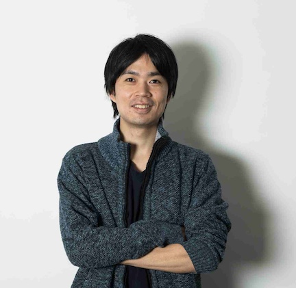
Dr. Tatsuya Osaki’s Biography:
Tatsuya Osaki is a Research Scientist at the Picower Institute for Learning and Memory at the Massachusetts Institute of Technology (MIT). He received his Ph.D. in Nano science and Nanotechnology from the University of Tsukuba in 2016. From 2016 to 2019, he worked as a postdoctoral fellow in the Department of Mechanical Engineering at MIT under the supervision of Prof. Roger D. Kamm. His research focused on developing microphysiological systems (MPS) to model tissue-tissue interactions, including the neuromuscular junction, blood-brain barrier, and neurovascular units, using microfluidic devices combined with optogenetics. His work aimed to elucidate physiological mechanisms and facilitate drug screening for neurodegenerative diseases such as amyotrophic lateral sclerosis (ALS). In 2019, he was appointed as a Project Assistant Professor at the University of Tokyo, Institute of Industrial Science, where he worked with Prof. Yoshiho Ikeuchi to advance microphysiological models for studying macroscopic neuronal circuits using interconnected organoid models. Since 2022, he has been a Research Scientist at MIT, working with Prof. Mriganka Sur. His current research focuses on investigating the pathological mechanisms of neurodevelopmental disorders using cerebral organoids and advanced bioimaging techniques. He utilizes three-photon microscopy to capture deep organoid neuronal activity and analyze alterations in neuronal dynamics in live organoids derived from patient-induced pluripotent stem cells (iPSCs).